Back Pain Doctor
Peroneal Tendinopathy
Written by Dr Joshua Hatch, Musculoskeletal GP, RACGP Fellow, Diploma in Musculoskeletal Medicine (FSEM UK).
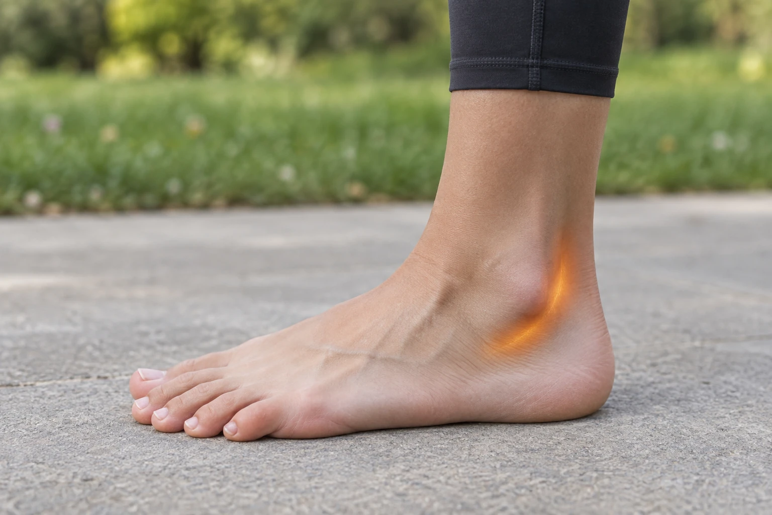
Peroneal tendinopathy affects the peroneus longus and peroneus brevis tendons, which curve behind the outer ankle bone on their way to the foot. They turn the sole outwards and are the first muscles to react when the ankle rolls, so they work hard in running and field sports and are commonly overloaded after ankle sprains. A high-arched foot with an inward-tilted heel increases the strain on them.
Assessment focuses on confirming the tendons as the source and looking for the reasons they are overloaded. That means examining the foot shape standing, testing the tendons and the ankle ligaments, and checking whether the tendons slip out of their groove. Ultrasound is used where a tear or subluxation is suspected. A sudden pop with the tendons flicking over the ankle bone is a different problem, an acute dislocation, which usually needs a surgical opinion.
First-line care is relative rest from the aggravating load, footwear or orthotic changes to unload the outer ankle, and a progressive physiotherapy programme covering tendon loading, balance and any ankle instability. A walking boot is used for a few weeks in more stubborn cases. The evidence for all treatment in this condition comes mainly from case series rather than trials, and there are no studies of shockwave for it. Surgery is considered when several months of well-run conservative care has not helped, or earlier for a significant tear or tendons that keep dislocating.
Common symptoms
- Pain and swelling behind and below the outer ankle bone
- Pain that builds with running, walking on uneven ground or cutting
- Pain when pushing the foot outwards against resistance
- Clicking or a feeling of the ankle giving way
Commonly seen in
Evidence for twelve treatment options in peroneal tendinopathy
This is not a fixed ladder or a package of treatments. Every option is assessed independently against this diagnosis, including treatments with limited direct research that may still be reasonable for a clearly defined tissue target. The evidence level, likely benefit, risks, cost and alternatives are weighed at consultation.
Foundation — diagnosis, education and rehabilitation
The part of care with the broadest relevance. For many people it is enough on its own; other options are judged against it, not automatically added after it.
Diagnosis, education and progressive rehabilitation Working out what is driving the pain, explaining what your imaging does and does not mean, and building a graded loading plan you can actually follow. First line, on case-series evidence
What it involves
Assessment to identify the likely pain source, an explanation of what the diagnosis means, adjustment of the loads that aggravate it, and a progressive strength and conditioning plan — with weight management where that is clinically relevant.
What the evidence shows
Non-operative care is the recognised starting point: relative rest and activity modification, anti-inflammatory medicines, orthoses with lateral posting, and a walking boot or cast for around six weeks in refractory cases, persisted with for four to six months before surgery is considered (StatPearls, Peroneal Tendon Syndromes 2023; Davda, EFORT Open Rev 2017). A specialist review notes that treatment recommendations for these disorders rest mainly on case series and expert opinion, and no randomised trial of a rehabilitation programme for peroneal tendinopathy was identified.
Pairing reviewed 2026-09-28.
Where it fits for you
Where I start: unloading the outer ankle, progressive peroneal and ankle strengthening, balance work, and footwear or orthotic changes. A high-arched, inward-tilted heel and lateral ankle instability are looked for at the first visit, because they are often the reason the tendons are overloaded.
Muscle and symptom-modifying options
Useful only when the examination identifies the tissue they target or when short-term pain control creates a useful rehabilitation window.
Wet needling and trigger point injection (trigger point injection) A fine needle into a tender band of muscle, with a small volume of dilute glucose solution injected through it. Only if relevant
What it involves
The needle is placed into the tender muscle band and a small volume of 5% glucose with 0.02-0.08% lignocaine is injected. Often done in the same visit as your assessment, and usable where the tender area sits deeper. Dry needling — the same technique with nothing injected — is not offered here.
What the evidence shows
A search pairing peroneal or fibularis tendinopathy with dry needling and trigger point treatment returned no study treating this condition with needling.
Pairing reviewed 2026-09-28.
Where it fits for you
Only where a separate tender band in the peroneal or calf muscle belly is reproducing part of the pain. It does nothing for the tendon itself.
Ultrasound-guided injections Guidance means we can see the needle reach the target. What goes into the needle depends on the diagnosis. Short relief for many
What it involves
The injection is performed under ultrasound so the needle can be watched into the target structure. What is injected is decided by the diagnosis — and corticosteroid is used sparingly, for the reasons set out below.
What the evidence shows
The largest series is 96 patients given an ultrasound-guided corticosteroid injection into the peroneal tendon sheath: 37 per cent reported more than 12 weeks of relief, 44 per cent a week or less, and a quarter went on to tendon surgery; shorter symptom duration predicted a longer response, and complications were sural nerve irritation and progression of one tear (Fram, Foot Ankle Int 2019). There is no controlled trial, and one specialist unit does not use steroid here at all because of the risk of tendon rupture (Davda, EFORT Open Rev 2017).
Pairing reviewed 2026-09-28.
Where it fits for you
Considered for sheath inflammation that has not settled with a proper rehabilitation trial, placed in the sheath under ultrasound rather than into the tendon, and with the rupture risk explained. It also helps confirm that the tendons are the source.
V-ACTOR vibration therapy (vibration therapy) Low-frequency vibration applied over tender muscle. Not a shockwave, despite sharing the machine. Plausible — no studies
What it involves
A vibration handpiece is moved over the tender muscle and fascia for a few minutes. It runs from the same STORZ MASTERPULS icon console as radial shockwave but is a separate handpiece delivering a different, gentler form of energy. No needles, no anaesthetic, no downtime.
What the evidence shows
A combined search for vibration therapy with peroneal tendon terms returned no study treating this condition.
Pairing reviewed 2026-09-28.
Where it fits for you
A comfort adjunct at most where calf or peroneal muscle guarding is limiting the exercise programme. Not a treatment for the tendon.
Energy-based treatment options
Radial pressure wave, focused shockwave and EMTT have different targets and different evidence. They are assessed separately rather than treated as one category.
Radial or focused shockwave? Read the overviewRadial shockwave (radial pressure wave) Broad treatment across tender muscle and fascia, close to the surface. Plausible — no studies
What it involves
A handpiece is moved over the tender tissue while ballistic pressure pulses are delivered, using a transmitter head chosen for the tissue — including heads shaped for fascia and for the muscle either side of the spine. The pulse is strongest at the skin and weakens with depth, so this is mainly a myofascial treatment, though it can reach deeper in some areas. No anaesthetic, no downtime, usually a course of sessions about a week apart.
What the evidence shows
A search for shockwave with peroneal or fibularis tendon terms returned two records: a single case report of a peroneus longus tear treated with focused shockwave alongside physiotherapy and orthotics (Culebras Almeida, Life 2026), and a case report of ankle ligament adhesions. No trial has tested radial shockwave in peroneal tendinopathy, and evidence from the Achilles or plantar fascia cannot be assumed to transfer.
Pairing reviewed 2026-09-28.
Where it fits for you
May be discussed for persistent tendinopathy after a full rehabilitation trial, on mechanism only and with the absence of evidence explained. Not used for a significant tear or for tendons slipping out of their groove, which are structural problems.
Focused shockwave (ESWT) A true shock wave converging at depth — for tendon, joint, ligament and bone. Plausible — no studies
What it involves
A focused system generates an acoustic shock wave that converges at a focal point set at a defined depth, concentrating the energy where the target is. This is the form we use for tendon, joint, ligament and bone. Typically four to six sessions about a week apart, no anaesthetic and no formal downtime.
What the evidence shows
The only published use is a single case report: a professional footballer with an isolated peroneus longus tear treated with eight sessions of ultrasound-guided focused shockwave plus physiotherapy and orthotics, who returned to play at 12 weeks (Culebras Almeida, Life 2026). A case report cannot separate the effect of shockwave from the rest of the programme.
Pairing reviewed 2026-09-28.
Where it fits for you
Same position as radial shockwave: discussed only after rehabilitation, as untested, and never in place of a surgical opinion for a tear or dislocation.
EMTT (magnetotransduction therapy) High-energy magnetic pulses applied over the area. Painless, no contact. Plausible — no studies
What it involves
You sit with the applicator positioned over the area for a short session. Painless, no gel or contact required, and usually delivered as a course — commonly alongside shockwave.
What the evidence shows
A combined search for electromagnetic and magnetotransduction therapy with peroneal tendon terms returned no study.
Pairing reviewed 2026-09-28.
Where it fits for you
Not part of managing this condition.
Biologic and joint injection options
Each injection is assessed independently for the named diagnosis. Cost, uncertainty, likely benefit, risks and alternatives are part of the decision.
Platelet-rich plasma (PRP) Your own blood, concentrated and injected under ultrasound guidance. Plausible — no studies
What it involves
Blood is taken from your arm, spun to concentrate the platelets, and injected into the target under ultrasound guidance in the same appointment.
What the evidence shows
The only data specific to these tendons are a subgroup of 23 peroneal patients within an uncontrolled pilot series of ultrasound-guided PRP for tendinopathy in general (Dallaudiere, J Vasc Interv Radiol 2014, cited in StatPearls). A 2023 review of non-operative management concludes that PRP is not currently supported by the evidence for peroneal tendon disorders (Curr Phys Med Rehabil Rep 2023).
Pairing reviewed 2026-09-28.
Where it fits for you
Not offered as a treatment for this condition on current evidence.
Prolotherapy (dextrose) A course of dextrose injections into and around the structure and its attachments. Plausible — no studies
What it involves
Hypertonic dextrose is injected at the tender ligament and tendon attachments, and into the joint where relevant, usually as a series of treatments a few weeks apart.
What the evidence shows
A search pairing peroneal tendon terms with prolotherapy and dextrose returned no study treating this condition.
Pairing reviewed 2026-09-28.
Where it fits for you
Not offered for this condition.
Viscosupplementation (hyaluronic acid) A gel-like injection into an arthritic joint. Widely used, and genuinely contested. Not used here
What it involves
One or a short series of hyaluronic acid injections into the joint under ultrasound guidance.
What the evidence shows
The only hyaluronic acid records with peroneal terms are laboratory and animal studies of a hyaluronic acid-gelatin coating to reduce adhesions on tendon grafts, not injection treatment of tendinopathy. There is no joint to inject.
Pairing reviewed 2026-09-28.
Where it fits for you
Not offered for this problem.
Nerve-targeted options
Relevant when examination identifies a superficial neuropathic pattern or a compressed peripheral nerve—not simply because pain is present.
Lyftogt perineural therapy (perineural injection therapy) A series of small dilute dextrose injections just under the skin, over tender superficial nerves. Only if relevant
What it involves
Small injections of low-concentration dextrose, usually 5%, placed just beneath the skin along tender superficial nerves. Given as a series of short sessions.
What the evidence shows
No trial of perineural dextrose in peroneal tendinopathy was identified. The sural nerve runs close to the tendons, and sural nerve irritation is a recognised complication of injections here (Fram, Foot Ankle Int 2019).
Pairing reviewed 2026-09-28.
Where it fits for you
Considered only if examination finds a separate superficial nerve pain pattern, such as sural nerve irritation. It is not treatment of the tendon.
Ultrasound-guided nerve hydrodissection Fluid placed around a nerve to free it from the surrounding tissue. Not a nerve problem
What it involves
Under ultrasound, fluid is placed in the plane around the nerve to separate it from the tissue that is tethering or compressing it.
What the evidence shows
The hydrodissection record that matches peroneal terms concerns the common peroneal nerve at the fibular head, a different structure at the knee (Song, Proc Bayl Univ Med Cent 2020). No study addresses the peroneal tendons, which are not a nerve entrapment.
Pairing reviewed 2026-09-28.
Where it fits for you
Only relevant if ultrasound and examination identify a separately compressed nerve, such as the sural or superficial peroneal nerve. Not a treatment for the tendons.
This is general information only and suitability is assessed individually. Treatments with limited or condition-dependent evidence may still be discussed, but only with clear explanation of the uncertainty, the expected benefit, risks, cost and alternatives. Red flags, progressive neurological symptoms or suspected serious pathology require a different pathway.
Every treatment above is described in full on the non-surgical treatments page, including what each one is used for and what the evidence supports. Ratings apply to the named diagnosis; an adjacent muscle, tendon, joint or nerve problem is assessed as a separate target. The method behind the ratings, and the treatments we do not offer, are set out on the evidence page.
Corticosteroid injection is reviewed separately for each diagnosis.
Cortisone reliably reduces pain in the short term, and that can be genuinely useful when pain is stopping you sleeping or moving enough to rehabilitate. Beyond that window the picture changes. A systematic review of 41 randomised trials and 2,672 participants found that corticosteroid injection reduced pain in the short term but that the effect reversed at intermediate and long term; in pooled analysis for lateral epicondylalgia the short-term effect was large (SMD 1.44, 95% CI 1.17 to 1.71) while no intervention was favoured at 26 weeks (-0.40, -0.67 to -0.14) and at 52 weeks (-0.31, -0.61 to -0.01) (Coombes, Lancet 2010). In a randomised trial in lateral epicondylalgia, 83 per cent of the corticosteroid group had completely recovered or much improved at one year against 96 per cent given a placebo injection, and recurrence ran at 54 per cent against 12 per cent; adding physiotherapy made no significant difference at one year (Coombes, JAMA 2013). In joints, repeated corticosteroid injections have been associated with cartilage loss. It does not repair anything.
So we do not build treatment plans around it. Where an injection is the right call, we review the specific diagnosis and target, discuss what is being injected and why, keep the number low, and pair it with the loading work that changes the longer-term course.
When we would still consider it
- Pain severe enough to block rehabilitation
- A clear inflammatory or bursal target on imaging
- A specific short-term goal — a trip, a procedure, a return to work
- Diagnostic use, where the response tells us where the pain is coming from
When a cortisone injection is worth doing, and when it is not
Other foot, ankle & lower leg conditions
Pain in the same area can come from more than one source, and the label matters less than what the examination and history point to. These are the other guides for this region.
Frequently asked questions
How is this different from a sprained ankle?
Do I need a scan?
Should I have a cortisone injection?
Ready for a clearer plan for your back or musculoskeletal pain?
Book an assessment with Dr Joshua Hatch.
Your assessment focuses on understanding the likely source of your pain and the most appropriate non-surgical options for your diagnosis, with the aim of reducing pain and improving function.
Book an appointment