Back Pain Doctor
Osteonecrosis of the Knee (Avascular Necrosis)
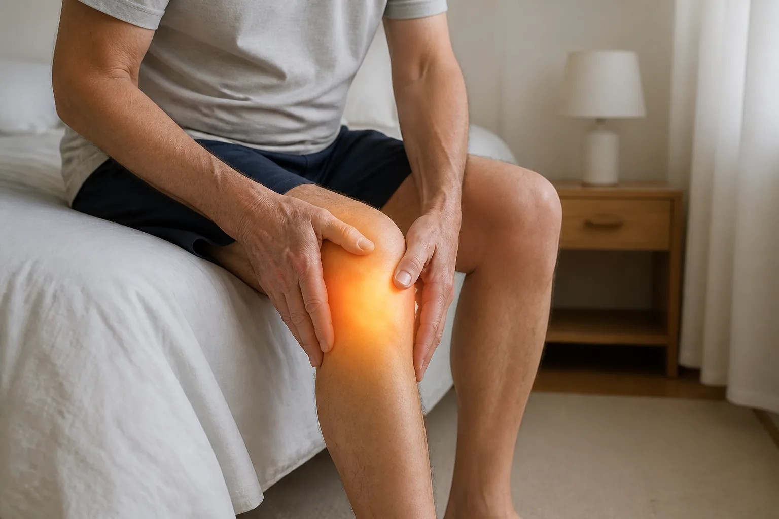
Osteonecrosis of the knee develops when the blood supply to a segment of bone — most often the medial femoral condyle on the inside of the knee — is disrupted. The affected bone dies and can collapse, and the overlying cartilage collapses with it, leading to arthritis of the joint.
Assessment focuses on distinguishing this from ordinary knee osteoarthritis, meniscal pathology and subchondral stress fracture. The history is often the clue: abrupt, well-localised medial knee pain in an older adult, or knee pain in someone on prolonged oral corticosteroids or with a transplant history. As with the hip, plain X-rays are usually normal early on, so ongoing symptoms with a normal X-ray warrant further assessment. MRI is the definitive investigation.
First-line care for small, early lesions is usually analgesia, protected weight-bearing, an unloader brace where appropriate, and a physiotherapy program to maintain quadriceps strength and joint range. Larger lesions, collapse, or pain that does not settle require orthopaedic assessment. The clinic’s regenerative and shockwave treatments do not have an established role in osteonecrosis, and the appropriate pathway here is timely diagnosis and specialist referral.
Common symptoms
- Sudden or well-localised pain on the inside of the knee
- Pain on standing and weight-bearing
- Swelling over the front and inside of the knee
- Tenderness to touch and reduced range of motion
Evidence for twelve treatment options in osteonecrosis of the knee (avascular necrosis)
This is not a fixed ladder or a package of treatments. Every option is assessed independently against this diagnosis, including treatments with limited direct research that may still be reasonable for a clearly defined tissue target. The evidence level, likely benefit, risks, cost and alternatives are weighed at consultation.
Foundation — diagnosis, education and rehabilitation
The part of care with the broadest relevance. For many people it is enough on its own; other options are judged against it, not automatically added after it.
Diagnosis, education and progressive rehabilitation Working out what is driving the pain, explaining what your imaging does and does not mean, and building a graded loading plan you can actually follow. Core care
What it involves
Assessment to identify the likely pain source, an explanation of what the diagnosis means, adjustment of the loads that aggravate it, and a progressive strength and conditioning plan — with weight management where that is clinically relevant.
What the evidence shows
Load is what builds bone; nothing else offered here does. A meta-analysis of 80 studies and 5,581 postmenopausal women found exercise improved bone mineral density at the lumbar spine, femoral neck and total hip, and that supervision was not required for the effect (Mohebbi, Osteoporos Int 2023). A network meta-analysis of 97 trials found every exercise type beat no exercise at the femoral neck, with resistance training ranking best for total hip (Zhang, J Clin Nurs 2021). Moderate-to-high impact loading also improves bone structure, not just density, and the effect is site-specific — it accrues where the load is applied (Ng, J Bone Miner Res 2023).
Pairing reviewed 2026-09-02.
Where it fits for you
For a bone stress injury the sequence is relative rest until it settles, then a graded return to load that is progressed slowly enough for the bone to keep up. For bone density, the loading is the treatment and it has to continue — the gains reverse when it stops. Energy availability, vitamin D and medical contributors are assessed alongside it.
Muscle and symptom-modifying options
Useful only when the examination identifies the tissue they target or when short-term pain control creates a useful rehabilitation window.
Wet needling and trigger point injection (trigger point injection) A fine needle into a tender band of muscle, with a small volume of dilute glucose solution injected through it. No trials in this condition
What it involves
The needle is placed into the tender muscle band and a small volume of 5% glucose with 0.02-0.08% lignocaine is injected. Often done in the same visit as your assessment, and usable where the tender area sits deeper. Dry needling — the same technique with nothing injected — is not offered here.
What the evidence shows
Searching osteonecrosis, avascular necrosis and bone marrow lesion against dry needling and trigger point injection returned three records, and all three are matches at other sites or on other treatments: jaw osteonecrosis after bisphosphonates (Almăşan, Rom J Morphol Embryol 2011), massage and acupressure for pain in sickle cell disease (Bodhise, Hematology 2004), and a facial pain case report (Adams, J Oral Pathol Med 1999). Nothing has tested needling in osteonecrosis of the knee.
Pairing reviewed 2026-09-19.
Where it fits for you
Not directed at the bone, which is where the problem is. Considered only where a separate tender muscle band is adding to your pain.
Ultrasound-guided injections Guidance means we can see the needle reach the target. What goes into the needle depends on the diagnosis. Only if relevant
What it involves
The injection is performed under ultrasound so the needle can be watched into the target structure. What is injected is decided by the diagnosis — and corticosteroid is used sparingly, for the reasons set out below.
What the evidence shows
Ultrasound improves needle-placement accuracy, but guidance is not a treatment in itself: benefit depends on a valid injectable target and what is injected. Condition reviewed: knee osteonecrosis.
Evidence source: Oo et al., ultrasound-guided injection systematic review (2024)
Pairing reviewed 2026-08-30.
Where it fits for you
For knee osteonecrosis, an ultrasound-guided injection is considered only when there is a specific diagnostic question or a separate joint, bursa, sheath or nerve target. The injectate and purpose are stated before treatment.
V-ACTOR vibration therapy (vibration therapy) Low-frequency vibration applied over tender muscle. Not a shockwave, despite sharing the machine. Not offered for this
What it involves
A vibration handpiece is moved over the tender muscle and fascia for a few minutes. It runs from the same STORZ MASTERPULS icon console as radial shockwave but is a separate handpiece delivering a different, gentler form of energy. No needles, no anaesthetic, no downtime.
What the evidence shows
Not appropriate over a systemic inflammatory process, healing bone, an area of osteonecrosis or a complete tendon or ligament rupture.
Pairing reviewed 2026-08-30.
Where it fits for you
Not offered for this.
Energy-based treatment options
Radial pressure wave, focused shockwave and EMTT have different targets and different evidence. They are assessed separately rather than treated as one category.
Radial or focused shockwave? Read the overviewRadial shockwave (radial pressure wave) Broad treatment across tender muscle and fascia, close to the surface. Not offered for this
What it involves
A handpiece is moved over the tender tissue while ballistic pressure pulses are delivered, using a transmitter head chosen for the tissue — including heads shaped for fascia and for the muscle either side of the spine. The pulse is strongest at the skin and weakens with depth, so this is mainly a myofascial treatment, though it can reach deeper in some areas. No anaesthetic, no downtime, usually a course of sessions about a week apart.
What the evidence shows
Shockwave reviews in myofascial pain report a possible benefit but mix protocols and often mix radial pressure wave with focused shockwave; that evidence cannot be transferred to the underlying diagnosis. Condition reviewed: knee osteonecrosis.
Evidence source: Wu et al., shockwave for myofascial pain review (2022)
Pairing reviewed 2026-08-30.
Where it fits for you
For knee osteonecrosis, radial pressure wave may be considered for a distinct superficial muscle or fascial component. It is not used as evidence of benefit for the primary diagnosis.
Focused shockwave (ESWT) A true shock wave converging at depth — for tendon, joint, ligament and bone. Worth discussing
What it involves
A focused system generates an acoustic shock wave that converges at a focal point set at a defined depth, concentrating the energy where the target is. This is the form we use for tendon, joint, ligament and bone. Typically four to six sessions about a week apart, no anaesthetic and no formal downtime.
What the evidence shows
Extrapolated from the femoral head, which is the site that has been studied (Zhang, Medicine 2017). No trial evidence at the knee specifically.
Pairing reviewed 2026-08-30.
Where it fits for you
Discussed with the indirectness stated, and alongside orthopaedic input rather than instead of it.
EMTT (magnetotransduction therapy) High-energy magnetic pulses applied over the area. Painless, no contact. Plausible — no studies
What it involves
You sit with the applicator positioned over the area for a short session. Painless, no gel or contact required, and usually delivered as a course — commonly alongside shockwave.
What the evidence shows
There are no published randomised trials of EMTT in this condition, or in any musculoskeletal condition, that we can point to. The supporting work is laboratory work: EMTT increased tenocyte proliferation, migration and collagen expression in cultured human tendon cells (Mancini, Int J Mol Sci 2025). The nearest human evidence for a related electromagnetic therapy is negative — a randomised trial of pulsed electromagnetic field therapy added to eccentric exercise for Achilles tendinopathy found no improvement in outcome scores or tendon neovascularity over eccentric exercise alone (Ko, Orthop J Sports Med 2024).
Pairing reviewed 2026-08-30.
Where it fits for you
Offered only as an adjunct alongside treatment that does have evidence, never as the plan itself, and only after that gap is explained. Much of the EMTT literature is manufacturer-affiliated, which is part of why we describe it this way.
Biologic and joint injection options
Each injection is assessed independently for the named diagnosis. Cost, uncertainty, likely benefit, risks and alternatives are part of the decision.
Platelet-rich plasma (PRP) Your own blood, concentrated and injected under ultrasound guidance. Only if relevant
What it involves
Blood is taken from your arm, spun to concentrate the platelets, and injected into the target under ultrasound guidance in the same appointment.
What the evidence shows
No role for PRP in the disease process here — a systemic, bone or pressure problem rather than a tendon or joint one.
Pairing reviewed 2026-08-30.
Where it fits for you
Considered only for a separate soft-tissue problem alongside it.
Prolotherapy (dextrose) A course of dextrose injections into and around the structure and its attachments. Only if relevant
What it involves
Hypertonic dextrose is injected at the tender ligament and tendon attachments, and into the joint where relevant, usually as a series of treatments a few weeks apart.
What the evidence shows
No role for prolotherapy in the disease process itself — this is a systemic, bone or structural problem rather than a painful ligament or tendon.
Pairing reviewed 2026-08-30.
Where it fits for you
Considered only for a separate mechanical problem alongside it.
Viscosupplementation (hyaluronic acid) A gel-like injection into an arthritic joint. Widely used, and genuinely contested. Only if relevant
What it involves
One or a short series of hyaluronic acid injections into the joint under ultrasound guidance.
What the evidence shows
No trial has tested hyaluronic acid in spontaneous osteonecrosis of the knee. Across the records it appears only as treatment for coexisting osteoarthritis or as salvage after something else failed: 11 of 74 people went on to viscosupplementation after percutaneous fixation for painful subchondral bone marrow oedema (DeBernardis, Arthrosc Sports Med Rehabil 2020), and a case report used it at four months for the osteoarthritis accompanying a subchondral insufficiency fracture (Gourlay, BMJ Case Rep 2015). The one randomised trial to measure bone marrow oedema after hyaluronic acid found it did not fall in the hyaluronic acid arm over twelve months, while it did where platelet-rich plasma was added (Zhang, J Clin Med 2025).
Pairing reviewed 2026-09-12.
Where it fits for you
Not a treatment for the bone lesion itself. Discussed only where the knee also has established osteoarthritis, and alongside load reduction and orthopaedic review.
Nerve-targeted options
Relevant when examination identifies a superficial neuropathic pattern or a compressed peripheral nerve—not simply because pain is present.
Lyftogt perineural therapy (perineural injection therapy) A series of small dilute dextrose injections just under the skin, over tender superficial nerves. Only if relevant
What it involves
Small injections of low-concentration dextrose, usually 5%, placed just beneath the skin along tender superficial nerves. Given as a series of short sessions.
What the evidence shows
Perineural dextrose evidence is concentrated in diagnosed peripheral compression neuropathies, particularly the upper limb; no direct evidence was identified for this diagnosis itself. Condition reviewed: knee osteonecrosis.
Evidence source: Perineural dextrose for upper-limb entrapment neuropathy review (2025)
Pairing reviewed 2026-08-30.
Where it fits for you
For knee osteonecrosis, perineural treatment may be considered only if a separate superficial neuropathic pain pattern is found on examination. It is not treatment of the primary diagnosis.
Ultrasound-guided nerve hydrodissection Fluid placed around a nerve to free it from the surrounding tissue. Only if relevant
What it involves
Under ultrasound, fluid is placed in the plane around the nerve to separate it from the tissue that is tethering or compressing it.
What the evidence shows
Hydrodissection evidence is concentrated in ultrasound-confirmed peripheral nerve entrapment, especially carpal tunnel syndrome; no direct evidence was identified for this diagnosis itself. Condition reviewed: knee osteonecrosis.
Evidence source: Buntragulpoontawee et al., hydrodissection injectate review (2021)
Pairing reviewed 2026-08-30.
Where it fits for you
For knee osteonecrosis, hydrodissection may be considered only if ultrasound and examination identify a separate compressed or tethered peripheral nerve. It is not treatment of the primary diagnosis.
This is general information only and suitability is assessed individually. Treatments with limited or condition-dependent evidence may still be discussed, but only with clear explanation of the uncertainty, the expected benefit, risks, cost and alternatives. Red flags, progressive neurological symptoms or suspected serious pathology require a different pathway.
Every treatment above is described in full on the non-surgical treatments page, including what each one is used for and what the evidence supports. Ratings apply to the named diagnosis; an adjacent muscle, tendon, joint or nerve problem is assessed as a separate target.
Corticosteroid injection is reviewed separately for each diagnosis.
Cortisone reliably reduces pain in the short term, and that can be genuinely useful when pain is stopping you sleeping or moving enough to rehabilitate. Beyond that window the picture changes. A systematic review of 41 randomised trials and 2,672 participants found that corticosteroid injection reduced pain in the short term but that the effect reversed at intermediate and long term; in pooled analysis for lateral epicondylalgia the short-term effect was large (SMD 1.44, 95% CI 1.17 to 1.71) while no intervention was favoured at 26 weeks (-0.40, -0.67 to -0.14) and at 52 weeks (-0.31, -0.61 to -0.01) (Coombes, Lancet 2010). In a randomised trial in lateral epicondylalgia, 83 per cent of the corticosteroid group had completely recovered or much improved at one year against 96 per cent given a placebo injection, and recurrence ran at 54 per cent against 12 per cent; adding physiotherapy made no significant difference at one year (Coombes, JAMA 2013). In joints, repeated corticosteroid injections have been associated with cartilage loss. It does not repair anything.
So we do not build treatment plans around it. Where an injection is the right call, we review the specific diagnosis and target, discuss what is being injected and why, keep the number low, and pair it with the loading work that changes the longer-term course.
When we would still consider it
- Pain severe enough to block rehabilitation
- A clear inflammatory or bursal target on imaging
- A specific short-term goal — a trip, a procedure, a return to work
- Diagnostic use, where the response tells us where the pain is coming from
When a cortisone injection is worth doing, and when it is not
Other knee conditions
Pain in the same area can come from more than one source, and the label matters less than what the examination and history point to. These are the other guides for this region.
Frequently asked questions
How is knee osteonecrosis diagnosed?
How is it different from knee osteoarthritis?
What does treatment involve?
Ready for a clearer plan for your back or musculoskeletal pain?
Book an assessment with Dr Joshua Hatch.
Your assessment focuses on understanding the likely source of your pain and the most appropriate non-surgical options for your diagnosis, with the aim of reducing pain and improving function.
Book an appointment