Meniscus Tear: Do You Need Surgery?
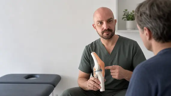
A meniscus tear does not automatically mean you need surgery. For many adults—particularly when symptoms developed gradually and the knee is not trul...


A Baker’s cyst does not automatically need draining. Many cause little or no trouble and can be observed while the knee problem producing the extra fluid is addressed. Drainage may be reasonable when a confirmed cyst is large, painful or restricts movement, but it is not a permanent fix and the swelling can return.
The important first step is to make sure the lump really is a Baker’s cyst, understand why the knee is making extra fluid and recognise symptoms that need urgent assessment.
A Baker’s cyst, also called a popliteal cyst, is a fluid-filled swelling at the back of the knee. It usually forms when excess joint fluid moves into a small sac between structures behind the knee.
In adults, the fluid is often associated with irritation inside the knee, such as knee osteoarthritis, inflammation or a meniscal problem. The cyst is therefore often a sign of what the knee joint is doing rather than a separate disease.
Some cysts are found by chance and cause no symptoms. Others create a feeling of fullness, tightness with deep bending or straightening, an ache after activity, or a visible lump behind the knee. The size of the lump does not always match how much it hurts.
Observation is often reasonable when the diagnosis is clear, symptoms are mild and the lump is not changing in a concerning way. Healthdirect notes that Baker’s cysts often settle without specific treatment.
The plan may focus on the knee rather than the cyst itself. Depending on the cause and the person, this can include:
Complete rest is rarely a useful long-term strategy. A graded plan aims to keep the knee functional without repeatedly provoking a large flare. No exercise programme can guarantee that a cyst will disappear, and an individual plan may be needed when the knee is very irritable or another diagnosis is present.
Aspiration uses a needle to remove fluid from the cyst, usually with ultrasound guidance so the target and nearby structures can be seen. In selected symptomatic cases, this may reduce pressure and pain for a period. A corticosteroid injection is sometimes considered at the same time or directed to the knee joint when inflammation is part of the picture.
The evidence needs to be kept in proportion. A 2020 systematic review found that image-guided injection and cyst fenestration had encouraging results, but the available treatment studies were low-level evidence and did not establish one best approach. A 2023 report of nine patients found meaningful symptom relief after image-guided aspiration and injection, but a small uncontrolled case series cannot predict how any one person will respond.
Drainage does not switch off the reason the knee made extra fluid. A cyst can refill, particularly if arthritis, inflammation or another joint problem remains active. That is why aspiration is better viewed as a symptom-management option for selected cases, not as proof that the underlying problem has been cured.
Surgery is uncommon. It may be considered when symptoms remain substantial despite reasonable non-surgical care, when the cyst repeatedly returns, or when a problem inside the knee needs an orthopaedic procedure for its own reasons. Removing a cyst simply because it appears on a scan is usually not the starting point.
A clinician may be able to recognise a typical Baker’s cyst from the history and examination. Ultrasound can confirm that a lump is fluid-filled, assess its features and help distinguish it from other causes of swelling. An X-ray may help assess arthritis, while MRI is sometimes used when the diagnosis is uncertain or an internal knee problem needs closer evaluation.
Imaging is particularly important when the lump is firm, growing, unusually positioned or does not behave like a typical cyst. Not every swelling behind the knee is a Baker’s cyst, so a new or changing lump should not be self-diagnosed from its location alone.
A Baker’s cyst can rupture and release fluid into the calf. This may cause sudden calf pain and swelling that can look very similar to a deep vein thrombosis (DVT). The symptoms alone cannot reliably tell the difference.
Seek urgent medical assessment for new or rapidly worsening one-sided calf swelling, warmth, redness or significant pain. If leg symptoms occur with chest pain, breathlessness, coughing blood, collapse or severe illness, call triple zero (000). Our guide to calf pain, muscle strain and blood clots explains why home tests cannot safely rule out DVT.
A hot, very swollen knee with fever or feeling unwell also needs prompt medical review. These features are not typical of an uncomplicated Baker’s cyst.
Most Baker’s cysts do not need automatic drainage. If the cyst is quiet, observation and management of the knee problem behind it may be enough. If it is painful, large or restricting movement, ultrasound-guided aspiration can be discussed with realistic expectations: it may reduce symptoms, but recurrence is possible and the supporting evidence is limited.
The practical takeaway is to treat the diagnosis and the person, not just the lump. Confirm what the swelling is, work out what is driving extra joint fluid, and seek urgent assessment if calf pain or swelling raises concern for a blood clot.
A meniscus tear does not automatically mean you need surgery. For many adults—particularly when symptoms developed gradually and the knee is not trul...
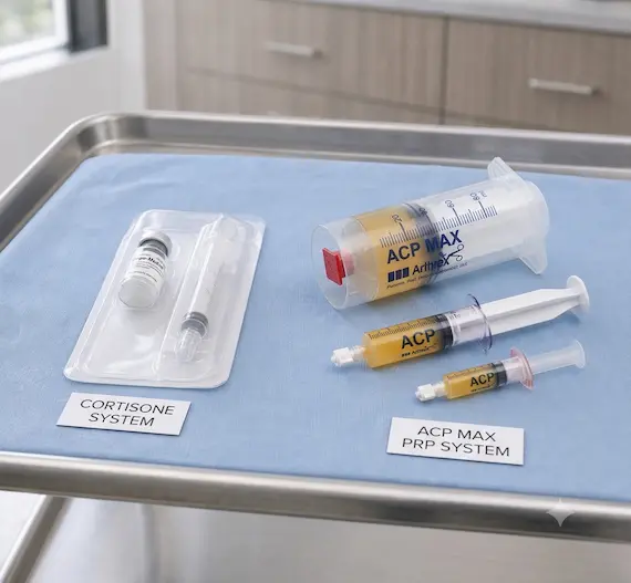
If you have knee osteoarthritis and have been offered an injection, you may be weighing up cortisone against PRP (platelet-rich plasma). They...

An injection is only useful if it reaches the right target. Some injections can be performed reliably using anatomical landmarks, but others are more...
Ready for a clearer plan for your back or musculoskeletal pain?
Your assessment focuses on understanding the likely source of your pain and the most appropriate non-surgical options for your diagnosis, with the aim of reducing pain and improving function.
Book an appointment